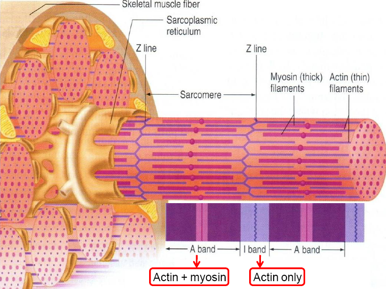
Skeletal Muscle Structure Pdf Actin Muscle Each myofibril contains 2 types of protein myofilaments. (myo = muscle) actin = thin myofilament myosin = thick myofilament collectively, these make up a functional muscle unit called a sarcomere. The document outlines the three types of muscles: skeletal (voluntary), smooth (involuntary, found in organs), and cardiac (involuntary). it details the structure and function of muscle proteins such as actin, myosin, nebulin, tropomyosin, and troponin, as well as the role of motor neurons and calcium in muscle contraction and relaxation.

Physiology Of Skeletal Muscle Contraction Pdf Thick myofilaments are composed of myosin. thin myofilaments are composed of actin. the organization of myofilaments produces the alternating light and dark striation characteristic of skeletal muscles. a sarcomere is a repeating pattern of a myofibril. myofibrils may be thought of as sarcomeres joined end to end. In this review, we discuss the various domains of muscle structure and function including its cytoskeletal architecture, excitation contraction coupling, energy metabolism, and force and power. Contraction occurs as a “muscle cell shortens”. what happens when skeletal muscle cells shortens? how do we explained muscle contraction using molecular biology? in this lecture we will focus on the structure and function of skeletal muscle cells. these cells are called muscle fibers. The muscle as a whole is surrounded by a sheet of dense connective tissues, called the epimysium. septa of this tissue extend inward, carrying the larger nerves, blood vessels, and lymphatics of the muscle. a single skeletal muscle is composed of numerous bundles of muscle fibers called fascicles.

Structure Of Skeletal Muscle Stock Vector Illustration Of Filaments Contraction occurs as a “muscle cell shortens”. what happens when skeletal muscle cells shortens? how do we explained muscle contraction using molecular biology? in this lecture we will focus on the structure and function of skeletal muscle cells. these cells are called muscle fibers. The muscle as a whole is surrounded by a sheet of dense connective tissues, called the epimysium. septa of this tissue extend inward, carrying the larger nerves, blood vessels, and lymphatics of the muscle. a single skeletal muscle is composed of numerous bundles of muscle fibers called fascicles. Skeletal muscles: well supplied with nerves and blood vessels. an artery and one or two veins accompany each nerve that penetrates a skeletal muscle. the neurons that stimulate skeletal muscle to contract are somatic motor neurons. microscopic blood vessels called capillaries are plentiful in muscular tissue;. Myofilaments are the three protein filaments of myofibrils in muscle cells. the main proteins involved are myosin, actin, and titin. myosin and actin are the contractile proteins and titin is an elastic protein. subject to conscious control. not under conscious control. The document outlines the ultramicroscopic structure of skeletal muscle, detailing components such as muscle fibers, myofibrils, sarcomeres, t tubules, and the sarcoplasmic reticulum.

Skeletal Muscle Howmed Skeletal muscles: well supplied with nerves and blood vessels. an artery and one or two veins accompany each nerve that penetrates a skeletal muscle. the neurons that stimulate skeletal muscle to contract are somatic motor neurons. microscopic blood vessels called capillaries are plentiful in muscular tissue;. Myofilaments are the three protein filaments of myofibrils in muscle cells. the main proteins involved are myosin, actin, and titin. myosin and actin are the contractile proteins and titin is an elastic protein. subject to conscious control. not under conscious control. The document outlines the ultramicroscopic structure of skeletal muscle, detailing components such as muscle fibers, myofibrils, sarcomeres, t tubules, and the sarcoplasmic reticulum.

Skeletal Muscle Pdf The document outlines the ultramicroscopic structure of skeletal muscle, detailing components such as muscle fibers, myofibrils, sarcomeres, t tubules, and the sarcoplasmic reticulum.