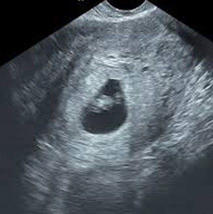
Youtube First trimester scanning is useful to identify abnormalities in the early development of a pregnancy, including miscarriage and ectopic pregnancy, and provides the most accurate dating of a pregnancy. first trimester scanning can be performed using either an abdominal approach or a vaginal approach. Sagittal: thalami midbrain, brainstem, 4th ventricle (intracranial lucency), and cisterna magna.

Pregnancy And Birth First Trimester Scan Nt Scan Southern Gem First trimester ultrasound (us) for anatomy assessment may improve anomaly detection but may also increase overall us utilization. we sought to assess the utility of first trimester us for evaluation of fetal anatomy. Ongoing technological advancements, including high frequency transvaginal scanning, have allowed the resolution of ultrasound imaging in the first trimester to evolve to a level at which early fetal development can be assessed and monitored in detail. The current goal of the first trimester ultrasound includes an element of fetal anatomic assessment and in experts’ hands, detailed evaluation of fetal anatomy is achievable and detection of several major fetal malformations is now possible with consistency. Describe the scanning protocol and techniques for a detailed first trimester ultrasound and compare the components of a standard versus detailed second trimester anatomy scan. summarize the clinical implications of common fetal ultrasound abnormalities and their utility by an interprofessional team.

First Trimester Fetal Morphology Scan Hkog Info The current goal of the first trimester ultrasound includes an element of fetal anatomic assessment and in experts’ hands, detailed evaluation of fetal anatomy is achievable and detection of several major fetal malformations is now possible with consistency. Describe the scanning protocol and techniques for a detailed first trimester ultrasound and compare the components of a standard versus detailed second trimester anatomy scan. summarize the clinical implications of common fetal ultrasound abnormalities and their utility by an interprofessional team. Summary of the structures recommended or suggested as part of the routine evaluation of the fetal anatomy in the first trimester, including the key features to check and the main anomalies potentially associated in case of abnormal features. First trimester ultrasonography (us) was first introduced for accurate dating of pregnancy based on the crown–rump length (crl) measurement and diagnosis of multiples. however, the rapid improvement in us imaging in the late 1980s proved that structural anomalies could already be detected in the first trimester. These guidelines represent an international benchmark for the first trimester fetal ultrasound scan, but consideration must be given to local circumstances and medical practices. We introduced a standardized anatomic protocol with 14 standard sections when performing routine first trimester ultrasound screening and reported its performance for detecting structural abnormalities in a large unselected population.